Investigating Chlamydomonas biosynthesis regulation
An excellent model organism in algae research
Dr Sandrine Bujaldon
Institut de Biologie Physico-Chimique
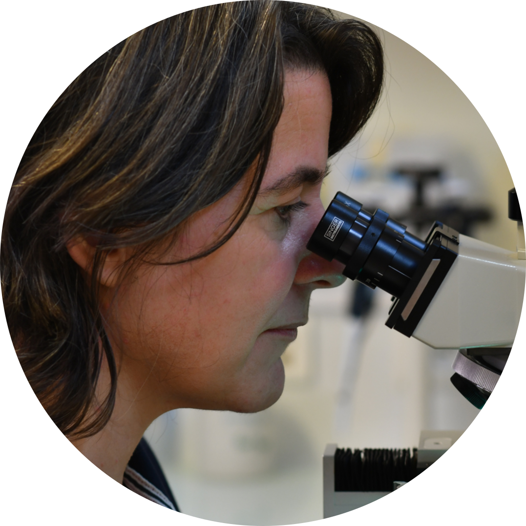
“Before using the MSM, I used to carry out tetrad dissections entirely manually, with a stereomicroscope. After I started with the MSM, there was no reason to look for another solution.”
Dr Sandrine Bujaldon
the Chloroplast Biology and Light Sensing in Microalgae laboratory
Institut de Biologie Physico-Chimique
Chlamydomonas Photosynthesis Research
Dr. Sandrine Bujaldon is a research engineer in the Chloroplast Biology and Light Sensing in Microalgae laboratory at the Institut de Biologie Physico-Chimique in Paris. She has dedicated her career to algae research, understanding the complex molecular processes that control how photosynthetic complexes are formed. She’s particularly focused on how Chlamydomonas reinhardtii, a green algae, adapts its photosynthesis to different light conditions – a crucial ability for optimising photosynthesis and ensuring survival.
By investigating these fundamental processes, Sandrine’s work not only deepens our understanding of Chlamydomonas photosynthesis at a molecular level, but also holds significant potential for real-world uses in agriculture and bioenergy. For example, her algae research could lead to developing crops with enhanced photosynthesis efficiency, allowing them to thrive in various light conditions. This could lead to increased crop yields and address global food security challenges.
Sandrine’s connection with Chlamydomonas began when 25 years ago she started her career managing a 500+ strain collection of photosynthesis mutants. This role involved extensive genetic crosses, and the use of the MSM became essential to her daily routine. Through this hands-on experience, Chlamydomonas became more than just a subject of study; it became a favored biological model, shaping her career and culminating in her recent PhD in biology, dedicated entirely to this fascinating organism.
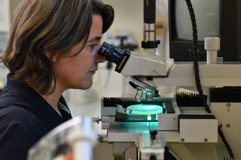
Photo credit: Gil Lefauconnier.
“Why is Chlamydomonas reinhardtii an excellent biological model?“
Through her early algae research work, Sandrine discovered the multitude of advantages that Chlamydomonas offers as a model organism. Its unicellularity simplifies complex molecular investigations into multicellular organisms. Meanwhile its amenability to culture and genetic manipulation has solidified its importance since the mid-20th century. Its versatility across diverse microbiological techniques, coupled with a rapid growth rate and a fully sequenced genome, facilitates in-depth analyses of various biological phenomena.
Particularly in photosynthesis and chloroplast function research, Chlamydomonas, with its similarities to plants, has proven invaluable. Its ability to grow heterotrophically, meaning without photosynthesis, while maintaining a functional photosynthetic apparatus, allows for the study of severe photosynthetic mutants. Furthermore, its a model organism for flagella structures analogous to cilia in human cells, helping us understand cellular movement and related pathologies. Finally, Chlamydomonas’ ability to produce hydrogen and its metabolic flexibility is incredibly promising for the biotech and bioenergy sectors.
The role of the MSM tetrad dissection microscope in algae research
“The use of MSM fulfills several research objectives linked to genetic analysis and strain manipulation.”
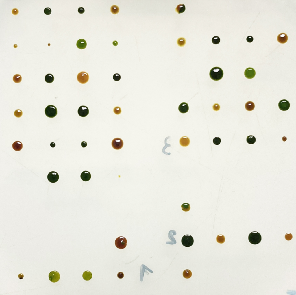
After many years of use, the MSM has helped Sandrine to precisely track and manipulate cells for thorough genetic studies, ensuring the reproducibility and accuracy of her algae research. Chlamydomonas tetrad dissection is a powerful tool for understanding how traits are inherited and for building custom genetic combinations, ultimately driving discoveries in fields like medicine and agriculture.
“Firstly, the MSM is essential for genetic segregation analysis. When a mutation is induced, whether spontaneously or resulting from mutagenesis, it is crucial to examine its mode of transmission. Next, the MSM is indispensable for purifying and cleaning Chlamydomonas strains. This step is essential to obtain homogeneous, well-characterised lines, thus guaranteeing the reliability of subsequent analyses.”
Dr Sandrine Bujaldon, Institut de Biologie Physico-Chimique
Sandrine’s most valued features of the MSM:
- Its optical system provides high clarity for observing delicate specimens, such as zygotes and tetrads.
- “A camera with a screen is indispensable for its educational value, learning micromanipulation is easier with this setup, making it highly beneficial for students’ training.”
- The comprehensive control afforded over the micromanipulator’s arms significantly enhances precision and operational flexibility. This makes the MSM comfortable and easy to use.
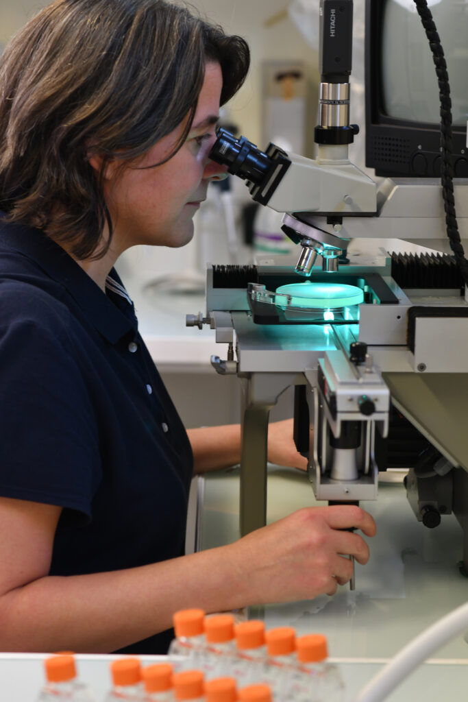
Photo credit: Gil Lefauconnier
Thousands of geneticists love the MSM
Listen to their stories here

Monica Perri | PhD Candidate
Monica Joined the UX and Research teams at Singer Instruments for her 3 month PIPS internship. She has been instrumental in shaping the development pipeline for all our products through detailed interviews with our users. Monica is studying at the University of Oxford investigating the evolution of low-oxygen sensing in early land plants.