High image quality
The key to accurate data and reliable results
Introduction
High-throughput lab automation is beginning to expand its reach within the Life Sciences, enabling labrats to get on with the fun stuff. However, what’s the point in having access to faster technology if the data it produces misses key insights?
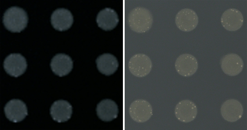
You can’t analyse what you can’t see… or can you? Same colonies – different colony imager.
Blurred lines:
How bad images ruin good experiments
Multiple key factors ensure that the images produced are not only visually clear but also true to the actual data they represent. A selection of these and the reason for their importance are listed below. When these are considered, the reasons why the control of your images is essential will become clear.
1. Flatness of light
An uneven distribution of light across the entire agar plate creates shadows and bright spots. If you were to then analyse colony areas across a plate, you would find an abundance of anomalies, making it challenging to accurately assess their true colours and intensities. Take these two images below for example, same plate but different imaging device, the difference is glaring especially after analysis.
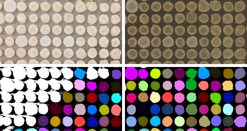
The same colonies before (top) and after (bottom) ImageJ colony analysis, using the same settings, but images from different cameras. The lighting is completely uncontrolled in the left image, but perfected in the right. The white particles were not detected by the analysis algorithm.
2. Colour
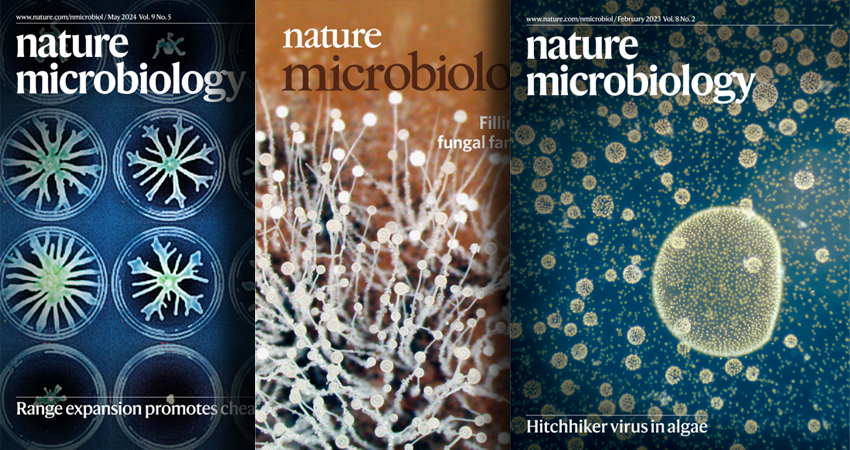
Colour fidelity (how accurately colours are represented in an image) and saturation (colour intensity) come hand-in-hand, enabling a scientist to distinguish between different colonies, reducing the risk of misinterpreting phenotypes or misidentifying a species. Without it you won’t ever have those striking images to grace the cover of Nature.
3. Resolution
Of course, a higher resolution and better contrast image allows you to see details you perhaps couldn’t capture before. Agar plates often contain colonies that vary in colour and texture, so high quality images allow for better differentiation between closely spaced colonies. If you study fine structures, this is a really important factor. There may be data there that you couldn’t have automated the interpretation of before.
Some colony wrinkles are tiny. Without high-resolution imaging, you just can’t see them.
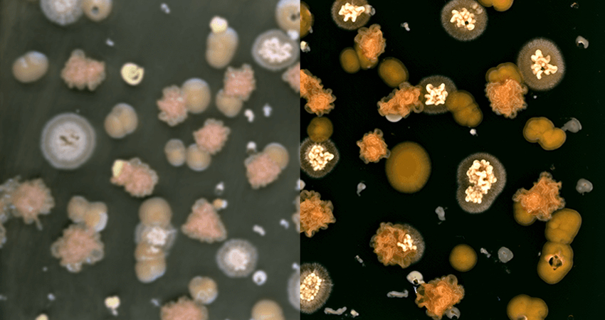
4. Saturation
An oversaturated image is one in which the intensity of certain pixels exceeds the optimal range, often due to excessive exposure or amplification of colour and brightness. This causes areas of the image to appear excessively bright or with exaggerated colors, leading to a loss of detail and making it difficult to distinguish between different elements in the image. This loss of detail will lead to an inaccurate image thresholding process (a common ImageJ analysis workflow) where important variations in the data are ignored, resulting in a misleading analysis.
Over-exposed Lion’s mane fungi (left) are never as sexy as the real deal.
The power of a clear image
In scientific research, clear and precise images are essential for accurate analysis and data interpretation, whether it’s observing colony phenotypes or documenting experiments. Image quality directly affects the accuracy and reliability of the information obtained from images (Bankhead, 2022). Poor quality data can lead to misinterpretations or errors, potentially compromising years of hard work. But, perhaps most importantly, you won’t ever have those sexy images on the cover of Nature. Images that will potentially propel your research to new heights and open up opportunities to share your discoveries with new audiences. Investing in high quality imaging equipment is therefore essential for promoting scientific success.
Quality at your convenience
Investing in high-quality is well worth the cost as it directly impacts the accuracy and reliability of your data. A better image leads to clearer observations, better data and clear conclusions. Your results will be more credible, your images sexier; publications will be clamouring to print your projects.
Looking for some insane image capture automation that’s better than your DSLR in a box?
Find the solution to all your image-acquisition problems with ColonyCam VOGUE.
Fiona Kemm MRes | Scientist
Fiona is a vital member of our Research team, rigorously testing our robots to ensure scientists don’t break them. With no prior robotics experience, she was the ideal guinea pig for our world-class user experience and support. Holding a BSc in Biochemistry and an MRes in Molecular Microbiology, Fiona brings extensive hands-on expertise she applies across departments, supporting both users and internal teams. From writing insightful web articles to specialising in SQWERTY, Fiona ensures our innovations perform flawlessly, helping customers focus on the creative and interpretive aspects of science that can’t be automated.