Single Cell Manipulation of yeast with the MSM 400 microscope
How scientists at the Max Planck Zentrum have harnessed the MSM 400 to select and screen individual yeast cells in array format.
Introduction
Dr Jona Kayser and his PhD student Nico Appold from the Max Planck Zentrum für Physik und Medizin are investigating evolutionary dynamics in dense yeast populations. By understanding how mutations arrive and survive in the compact colony community, Jona models how anti-microbial resistance and cancer develops in infections and tumours respectively.
This work has led to development of a powerful new experimental evolution approach. Nico developed a yeast strain that undergoes predetermined, inheritable and irreversible mutations which cause changes in fluorescence and resistance to temperature treatment. Their platform can track the entire evolutionary path of thousands of individual yeast cells to better understand how resistant cells take hold (Aif et. al., Nature 2022). Microfluidic experiments have further extended this work showing the importance of spatial factors, like cellular crowding, for the emergence of rare genetic variants (Schreck et. al., PNAS 2023).
Building on these findings, Nico is leading a study to better understand evolutionary dynamics at the cellular level. Using the MSM 400, he plans to microscopically array non-resistant single cells in 20-point grids to observe how mutations emerge and how treatment influences their evolution. Understanding the evolutionary dynamics and development of treatment resistance in pathogens could influence how anti-microbial resistance is combated in the future.
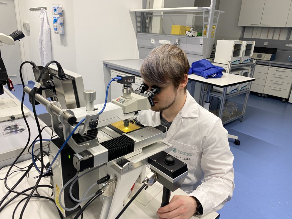
Figure 1: Nico Appold using the MSM 400 to select single cells to create 20-point arrays on SBS format agar plates.
Methods
This method is for the separation of individual cells from liquid culture and the placement of these cells to create a 20 array of clonal colonies.
Yeast inoculation:
- Prepare a liquid culture of yeast at least 4 hours prior to seeding and incubate at the optimum temperature.
- The culture OD600 should be less than 1 after incubation. Dilute with additional broth if necessary.
- Transfer 1 ml of culture into an Eppendorf tube and vortex to separate the yeast clusters.
- With 10-50 µl of culture make a broad streak across “column 1” of an agar PlusPlate (Figure 2).
- Use a sterilised glass spatula to spread the streaked culture across the area from “column 1” to the nearest plate edge.
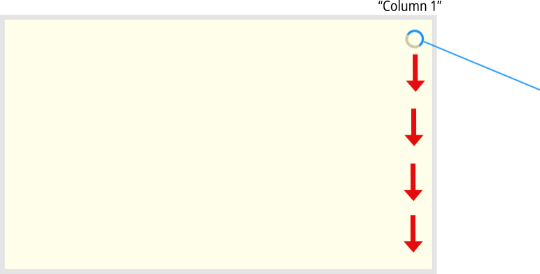
Figure 2: Streak the culture down the right hand side of the agar plate. When the plate is inverted on the MSM 400 the streak will be in “column 1” on the left hand side.
MSM 400 set up:
- Lift head of the MSM 400 microscope. Adjust needle to the appropriate height for the agar depth and remove the petri dish spacers (black screws in the sample holder).
- Carefully place the PlusPlate with the inoculum on the left hand-side.
- Choose the PlusPlate viewer option from the menu and select the grid you require.
- Use the joystick on the left to move the plate so that the needle is visible through the inoculum streak and press the black button.
- This sets the position as A1 and will allow you to access your yeast by returning to any position in column 1.
- Click on the menu button in the bottom right of the touchscreen, select “Layout” and pick one where the grid and camera picture are both displayed.
- This allows you to easily switch between the grid and the inoculum by using the touchscreen (Figure 3).
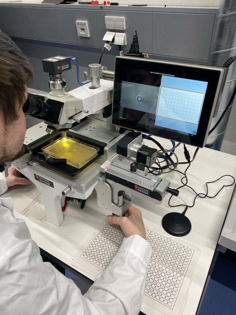
Figure 3: Using the MSM 400 to pick up and place single cells is easy to learn and a simple solution to semi-automating single cell manipulation.
Single cell manipulation:
- Select a single yeast cell that looks alive from the inoculum and take it onto the dissection needle.
- The best cells have a small bud.
- Use the Zapper to separate any large clusters
- Use the touchscreen to select the desired target position on the grid and place the cell.
- Placing the cells on every other position of a 96 array ensures there is enough space and nutrients for long term experiments.
- A diagram of the appropriate array structure can be seen in Figure 4.
- Return to the inoculum by selecting a position in column 1 using the touchscreen (Figure 5).
- Repeat this process until you have transferred all the desired cells.
- Incubate the plate and observe the population growth from single cell to clonal colony.
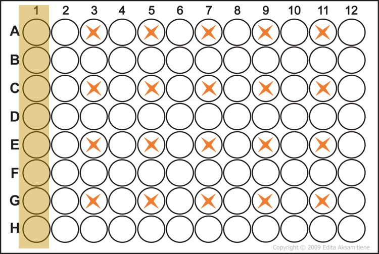
Figure 4: The inoculation schematic of the agar PlusPlate. Single cells are placed at every other position (*) from the source streak of culture. This diagram is from the perspective of the MSM 400 with the agar facing downwards.
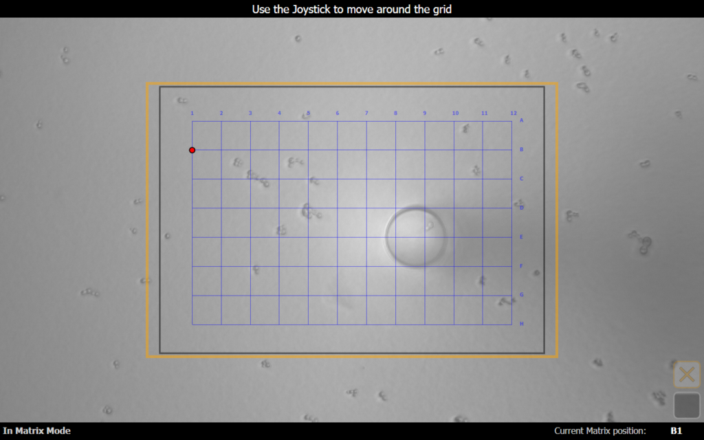
Figure 5: Nico picks a healthy individual cell from column 1 with the needle. The MSM 400 software displays the 96 grid overlaid with the red dot indicating the current position viewed in the background
Results
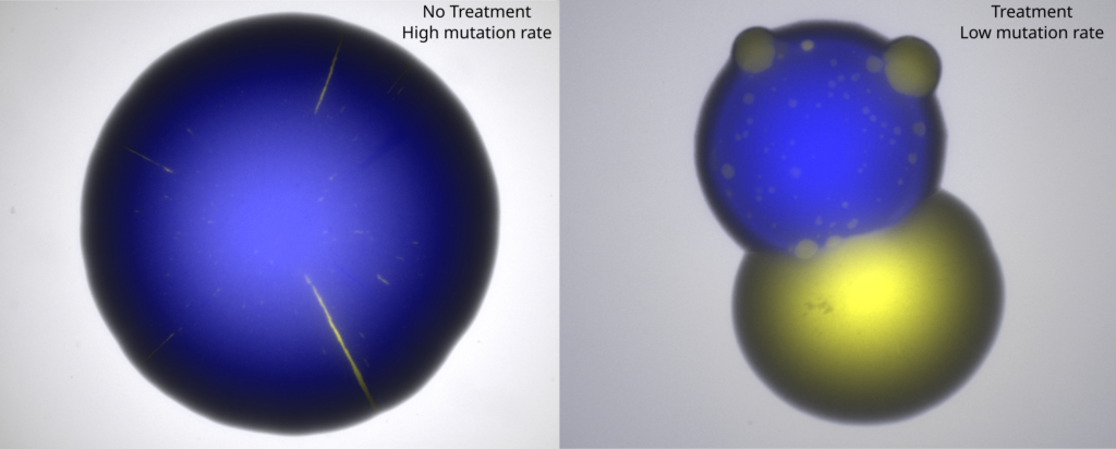
Figure 6: Colonies 75 hours after inoculation by the MSM 400. The results demonstrate that treatment inhibits the growth of blue fluorescent cells, whereas resistant yellow fluorescent cells continue to proliferate.
After the array of yeast was inoculated by the MSM 400 Nico adjusted the mutation rate of some cells to be high and others low. The plates were then subject to treatment to track how resistance to said treatment developed. Figure 6 demonstrates that the treatment inhibited the growth of blue fluorescent cells (right blue colony is far smaller than left) but the yellow fluorescent cells continued to proliferate. The yellow fluorescence indicated the introduction of mutations which caused their resistance to the treatment. Thus the yellow fluorescent cells continued to proliferate in spite of the treatment, in other words “treatment failure”.
“The MSM 400 has been instrumental in increasing the throughput and control-ability of our experiments”
Nico Appold, Max Planck Zentrum für Physik und Medizin
Thank you Nico and Jona for sharing this unique workflow for precision control over single cell selection with the MSM 400. Single cell manipulation from under a microscope will allow any yeast scientist to skip the streaking and clone isolation steps to create arrays of clonal colonies in hours rather than days. No need for an automated cell sorting system.
This low throughput semi-automated solution to creating arrays is another reason why the MSM 400 is the perfect tool for manipulation and observation of single cells.
Unlock the secrets of single cell behaviour with the MSM 400!
Track the changes in single cells through automation.

Fiona Kemm MRes | Scientist
Fiona is a vital member of our Research team, rigorously testing our robots to ensure scientists don’t break them. With no prior robotics experience, she was the ideal guinea pig for our world-class user experience and support. Holding a BSc in Biochemistry and an MRes in Molecular Microbiology, Fiona brings extensive hands-on expertise she applies across departments, supporting both users and internal teams. From writing insightful web articles to specialising in SQWERTY, Fiona ensures our innovations perform flawlessly, helping customers focus on the creative and interpretive aspects of science that can’t be automated.