Xenopus Embryo Microinjection
How the National Xenopus Resource uses an MK1 micromanipulator to inject hundreds of frog embryos a day
By Esther Pearl and Marko Horb
Marine Biological Laboratory, University of Chicago
Microinjection is an important technique used by many Xenopus researchers to inject into embryos for loss-of-function1 and gain-of-function studies 2,3, making transgenic 4,5,6 and knock outlines 7, as well as injecting into oocytes 8,9,10.
The National Xenopus Resource (NXR), based in Woods Hole, Massachusetts, USA, acts as a resource center for Xenopus research, offering custom-made transgenic frogs, TALEN and CRISPR knock-out lines, as well as maintaining existing frog lines. We also perform oocyte host transfer (transferring oocytes that have been injected with mRNA into a host female to be laid and then fertilized), all with the help of the MK1.
A particular benefit of the MK1 is it allows us to microinject Xenopus oocytes and embryos using natural hand movements, as opposed to using knobs. Thus it is very intuitive and your hand never has to leave the handgrip.
The MK1 moves the needle in the same direction as the hand moves, only the movement is reduced by a 1:4 ratio. This allows fine control of the injection needle, enabling precise injections. The MK1 can be rotated so that the needle is parallel to the bench, or on an angle, without the need for an additional tilt mount.
We routinely inject into Xenopus embryos and oocytes at various stages, including 1-cell, 2-cell, 8-cell and up to 32-cell stages. This allows us to target specific areas of the developing embryo if necessary. Using the MK1 makes it much easier to control the area of the embryo we are injecting.
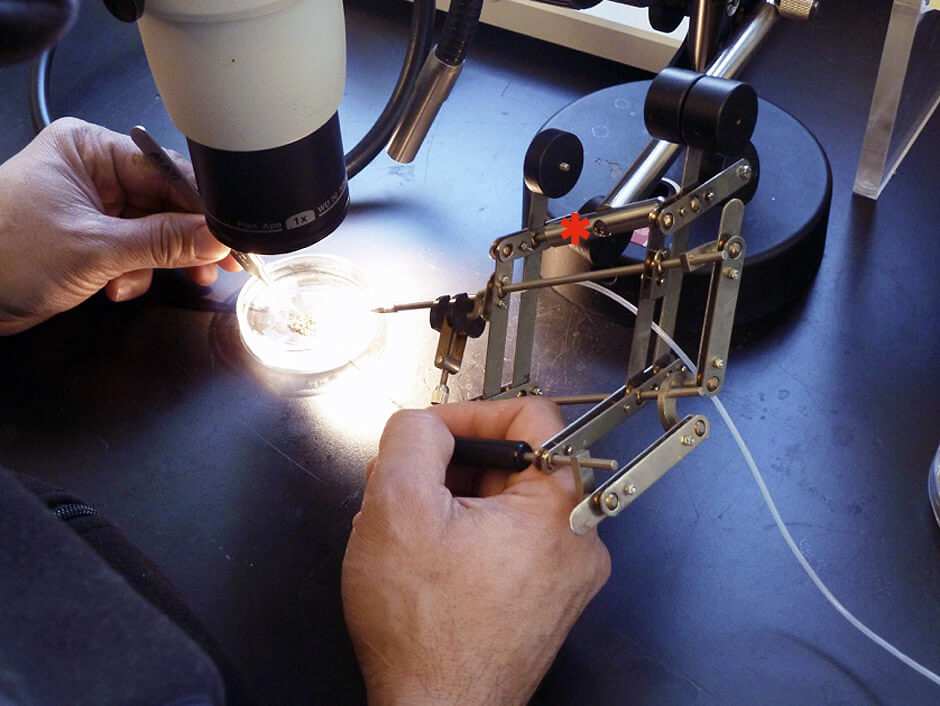
Some of our injections are into the vegetal pole and use of the MK1 allows for precise injection into the vegetal hemisphere, which is the heavier side of a dejellied embryo, and thus on the bottom. The angle of the pipette holder can easily be adjusted at any time throughout the injection procedure, by tilting the bar where the pantograph meets the horizontal bar connecting to the stand (see asterisk in Figure 1).
Being able to use natural hand movements enables new users to adapt to and use the micromanipulator effectively very quickly. It also has an ergonomic design that reduces muscle fatigue, which in turn increases efficiency enabling us to inject more embryos in a day. The NXR regularly injects hundreds, often thousands, of embryos a day; the MK1 enables us to perform efficiently.
Intrigued to find out what else the MK1 can do?
Expertly designed for delicate, steady handling and control of precision needles and tools.